The pertinent anatomy is described and the role of the tympanic diaphragm and isthmus in determining the degree to which middle ear disease may progress is stressed.
Attic retraction ct.
Five years after the initial examination otoscopic examination revealed an intact tympanic membrane with a tiny attic retraction fig.
In 26 of 28 ears with attic retraction pockets at least a portion of attic was aerated and in 22 of these 26 ears.
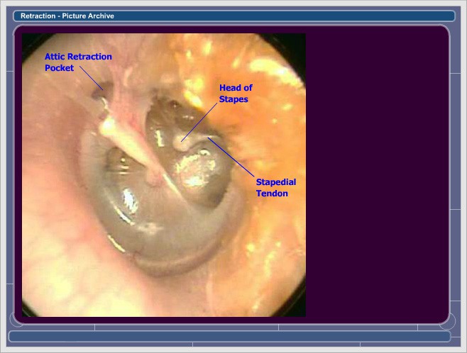
Tympanic membrane retraction usually occurs when a portion of the tympanic membrane becomes weakened and is pulled inwards by the negative pressure within the middle ear.
The appearances on ct scans of chronic otomastoiditis tympanosclerosis cholesterol granuloma attic retraction pocket and acquired cholesteatoma are reviewed and illustrated.
The retraction can be subdivided based on severity 1.
An acquired attic cholesteatoma may spontaneously drain externally into the external auditory canal leaving a cavity in the attic with the shape of the original cholesteatoma but now filled with air a phenomenon referred to as nature s atticotomy or auto atticotomy.
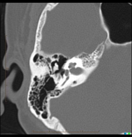
Ct is the modality of choice for diagnostic assessment of cholesteatomas due to its ability to demonstrate the bony anatomy of the temporal bone in exquisite detail.
To examine this theory computerized tomographic ct findings of these conditions were evaluated in a series of 53 ears with retractions of the pars flaccida attic retractions of tos type ii or deeper including both retraction pockets and cholesteatomas.
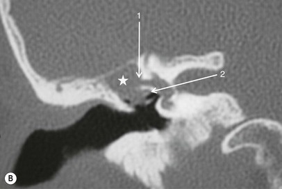
Attic retraction pocket in the left ear white arrow with atelectatic prussak s space red circle and eroded scutum yellow arrow.
We describe and quantify the ct appearance of the auto atticotomy cavity as it pertains to the.
In contrast in the 25 cases with attic cholesteatomas these numbers decreased to 10 and 5 respectively and the lack of aeration of the attic was demonstrated in 15 of 25 60 of the cases.
Cholesteatomas appear as regions of soft tissue attenuation exerting mass effect and resulting in bony erosion.
2c and a follow up temporal bone ct scan showed a soft tissue density lesion in the lateral and superior portions of the incudomallear joint fig.
Download citation a tiny retraction of the pars flaccida may conceal an attic cholesteatoma objective the purpose of this study was to evaluate the possibility of attic cholesteatomas.
In 26 of 28 ears with attic retraction pockets at least a portion of attic was aerated and in 22 of these 26 ears the mastoid antrum was also aerated.